If you’ve ever visited a dentist, chances are you’re familiar with a dental X-ray. The most common are bitewing X-rays, which detect tooth decay, dental tartar and gum disease. But there are actually several types of dental X-ray images used to diagnose dental problems that aren’t visible to the naked eye. One of these is called a cephalometric X-ray, or a CEPH.
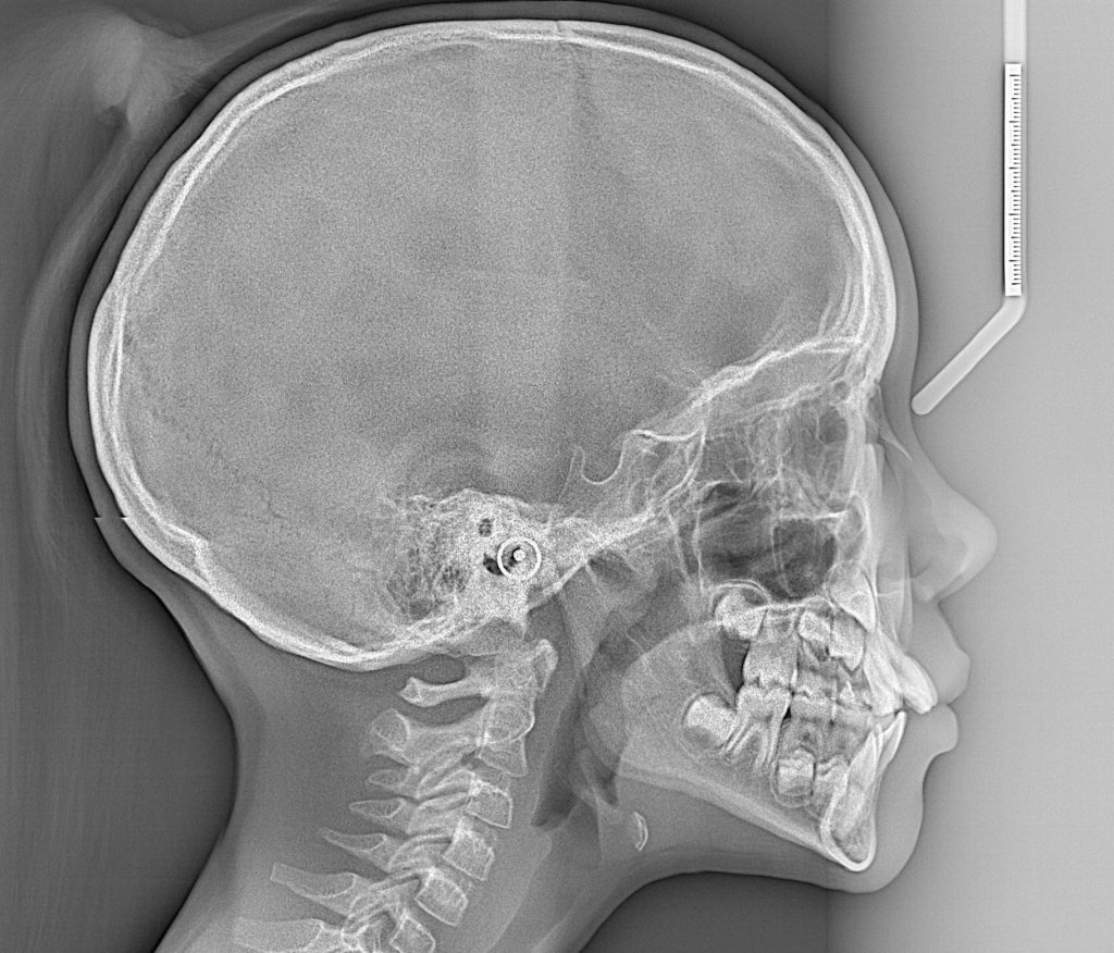
CEPH X-ray pictures capture images of the entire skull and profile. CEPH is most often used by orthodontists to diagnose problems with your jaw and bite that might improve with orthodontic tools such as dental braces. This type of dental X-ray also allows the dentist to measure irregularities in your facial profile.
If your dentist or orthodontist recommended that you have a CEPH, you might be wondering how it can help and exactly what you can expect from the dental X-ray experience. Here’s some information we hope will help answer your questions.
CEPH X-rays at Work
CEPH is an extraoral X-ray, which means the film is outside the mouth. These types of images look for impacted teeth, monitor growth and development of the jaws in relation to the teeth, and identify potential problems between teeth and jaws and the temporomandibular joint (TMJ) or other bones of the face. The CEPH X-ray is used in the following areas of dental care:
Orthodontic Planning — An orthodontist uses CEPH X-ray pictures to diagnose misalignment of the jaw and bite problems. A CEPH X-ray is used primarily for treatment planning. Once the dental X-ray image is taken, the dentist can trace the CEPH to calculate how your jaw and surrounding bone will be affected by dental treatment.
Sleep Apnea Treatment — CEPH X ray pictures of the skull provide a view of a person’s airways. This makes them helpful in the diagnosis of problems such as sleep apnea, a disorder in which people literally stop breathing during their sleep.
The CEPH X-ray Process
Taking a CEPH X-ray is very similar to taking a Panorex X-ray, which is a single picture of all your teeth and surrounding bones. CEPH X-ray pictures are taken on a standard panoramic machine equipped with a cephalometric film-holding arm mounted off to one side. Many dental offices have the machinery and equipment necessary to take CEPH X-ray pictures.
During the X-ray, you will wear a lead apron to protect you from radiation and the technician will position you as needed inside the dental X-ray machine. The process is quick — about 10 seconds — and the image is developed in less than 10 minutes. Best part? It’s totally painless!
As with other types of X-ray pictures, CEPH can be taken on conventional radiographic film or it can be developed using digital radiography. Digital images are quicker, use less radiation and can be enhanced, enlarged and shared via computer.